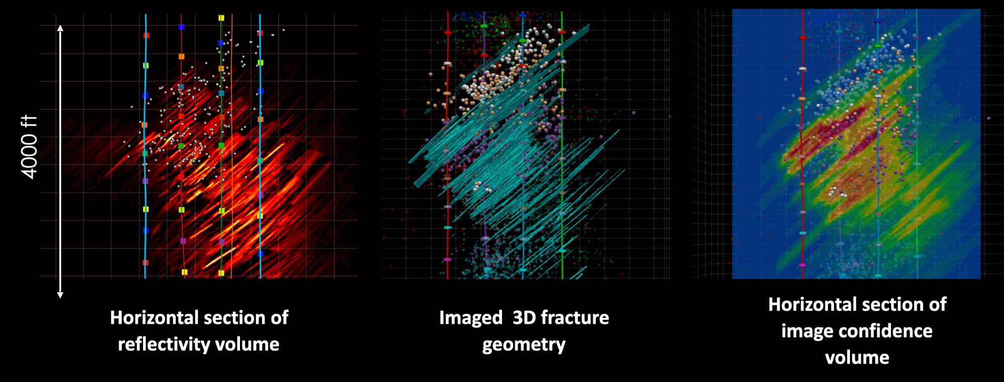
Trademark
Trademark

Method Overview

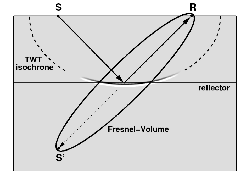
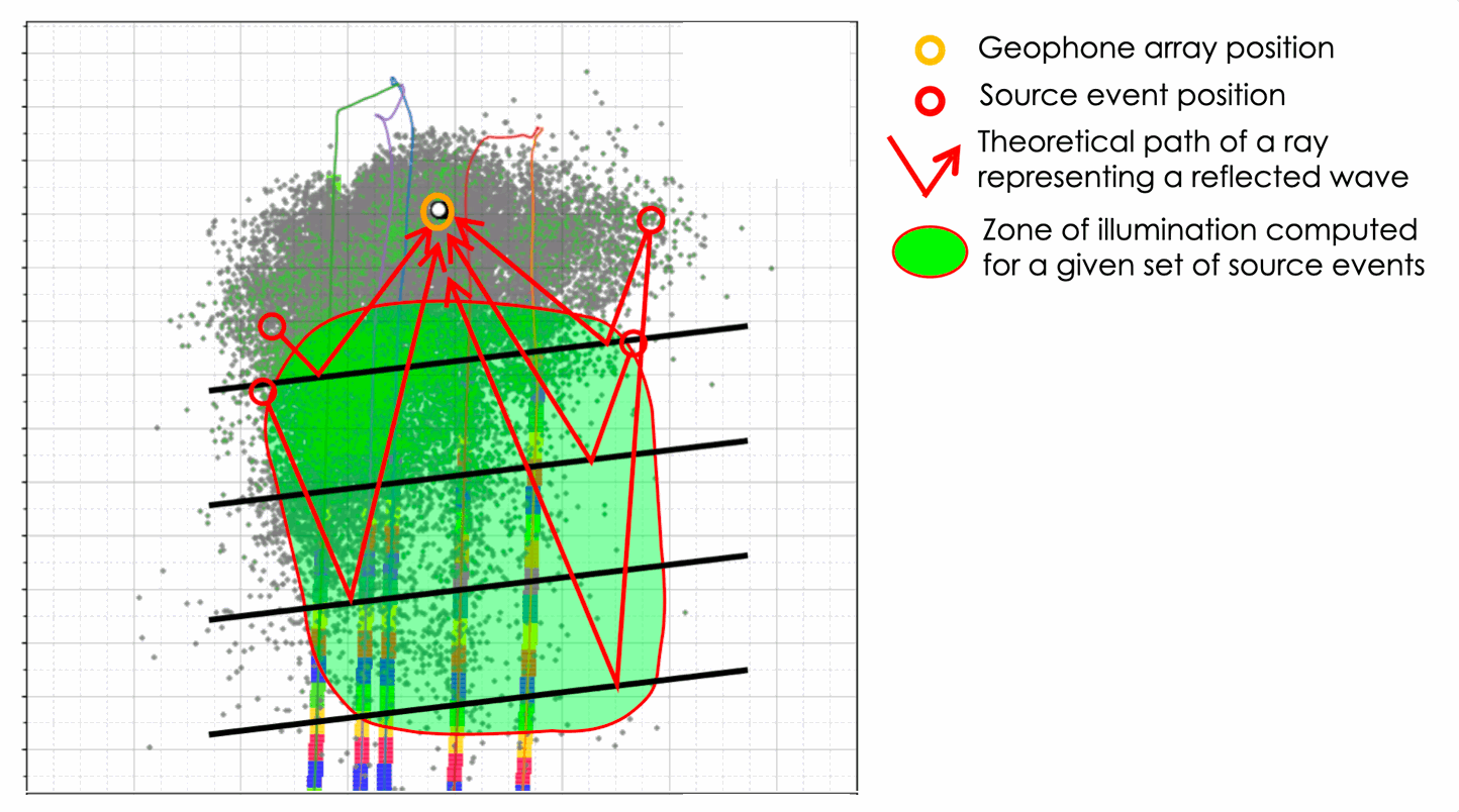
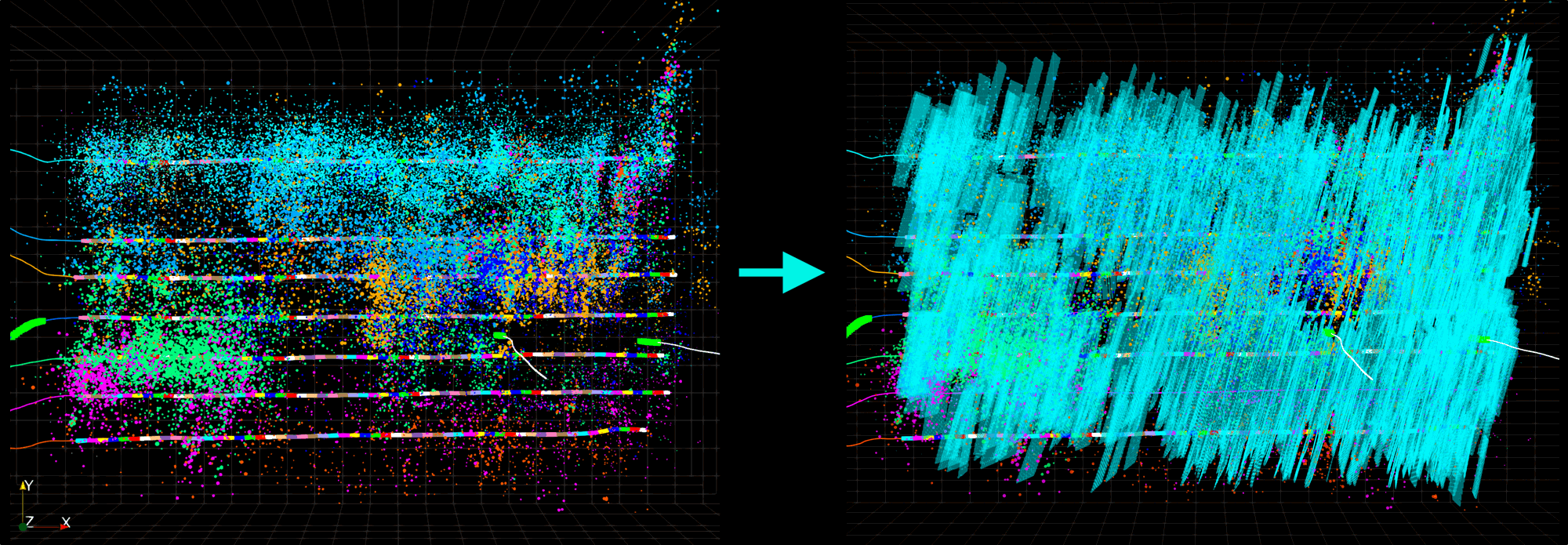
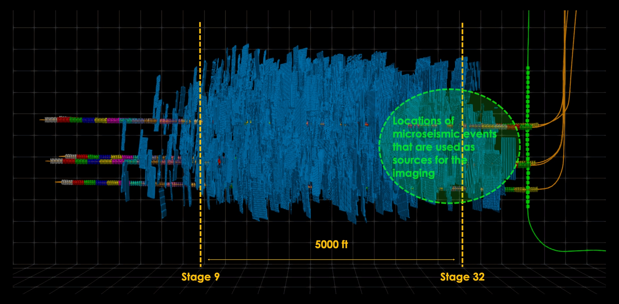
Method uses microseismic events as sources for reflection imaging to image newly created and pre-existing fractures.The algorithm is designed to scan microseismic waveform data and extract a high number of potential reflections. Multiple reflections created by the same fracture network recorded by a fixed array of sensors which helps to overcome the uncertainty of individual event localizations.
Fracture Imaging Technology Overview

Advantages of Microseismic Events as Sources

Large number of source events
Sources are located within the area of interest
High frequency signals → high resolution of the final image

Reflection Imaging Algorithm

To handle low aperture microsiesmic data we use a custom imaging operator that is based on Fresnel Volume Imaging algorithm. The operator is constrained to the area around the actual reflection point either by using polarization of 3C downhole data or by FI fracture orientation scanning in case of 1C DAS data.

Imaging Workflow:

Scan waveform data and extract potential reflections
Use custom imaging operator to compute location, size and orientation of all potential reflection surfaces
Compute stack in 3D reflectivity volume along with FI confidence volume
Extract 3D reflector geometry representing the strongest reflectors within the reflectivity volume
Fracture Imaging: imaging operator
Source: Lüth et al. (2005), Buske et al. (2009)

Zone of Illumination

The zone of illumination is computed theoretically using locations of sources and receivers. It is based on the assumption that reflectors (fractures) are aligned along ShMax.
In order to compute this zone we take events on the edges of a microseismic cloud and compute maximal possible area that can be illuminated from these edge events using a given acquisition system.
The theoretically computed zone of illumination does not represent extend of the final image. It shows the extend of the area for which reflections from optimally oriented fractures can be captured by a given downhole sensor/DAS array(s).
Fracture Imaging: Zone of Illumination

Application and Outputs

Applicability

Compatible with acoustic data from borehole geophones or any fiber DAS.
Adds value to both legacy and new datasets — no special acquisition setup or extra costs required.
Fracture Imaging is a mature technology, analytically validated against other diagnostics including: Cross-Well-Strain, Permanent DAS in-well production, Time-lapse RFS-DSS production profiling, Core, & 3D Seismic.

Deliverables

Fracture Geometry: Provided as a 3D reflectivity volume and a set of triangulated surfaces.
Fracture Morphology: Detailed characterization from near-wellbore to far-field, including aseismic zones, natural faults, and interactions with depleted wells.
Fracture Surface Area and Relative Conductivity: Quantified and segmented by well or completion design group.
Perforation cluster perfomance metrics: Includes cluster uniformity and bias.
Fracture Imaging Analytics: A suite of reproducible metrics defined by project objectives. Examples are available on the Analytics page.
Fracture Imaging Technology: Input and Output

Far-Field Frcture Imaging

Fracture Imaging technology can image fracture geometry up to ~5,000 ft away from the microseismic events used as sources. This long-range imaging is achievable even with data recorded by standard borehole geophones, provided the number of source events is sufficiently high (typically 5,000–10,000).
Far-Field Fracture Imaging

Fracture Imaging from DAS data

Fracture Imaging is fully applicable to microseismic data recorded by DAS arrays, even though such recordings lack polarization information. This absence introduces ambiguity in locating reflectors. To resolve this, we apply a set of physically grounded assumptions—most importantly, that reflections predominantly originate from sub-vertical fractures.
To estimate fracture orientation, we perform an imaging scan across a range of possible fracture azimuths. The orientation that yields the highest stack quality is selected, based on the principle that correct alignment produces constructive interference and a more coherent image.
Fracture Imaging using DAS data